-
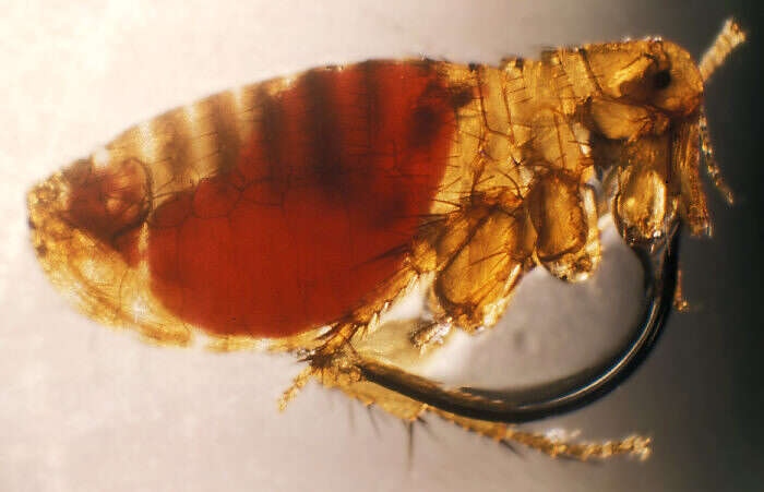
Scanning Electron Micrograph of a Flea. See PHIL 11436 for a colorized version of this image.Created:
-
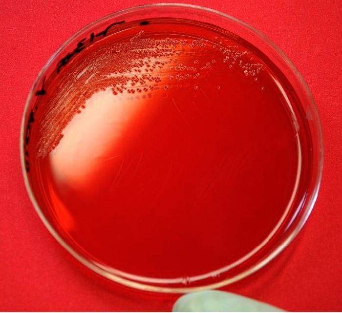
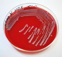
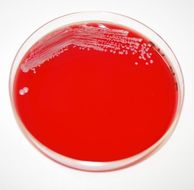
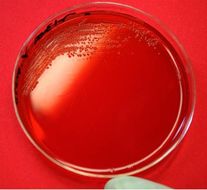
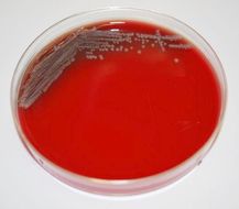
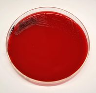
This image depicts a Petri dish containing a sheeps blood agar (SBA) medium, which had been inoculated with Gram-negative Yersinia pestis bacteria. Y. pestis is the pathogen responsible for causing human plague. This was the appearance of the colonial growth after 96 hours of incubation at 25º C.People usually get plague from being bitten by a rodent flea that is carrying the plague bacterium or by handling an infected animal. Millions of people in Europe died from plague in the Middle Ages, when human homes and places of work were inhabited by flea-infested rats. Today, modern antibiotics are effective against plague, but if an infected person is not treated promptly, the disease is likely to cause illness or death.Created: 2009
-
This image depicts a Petri dish containing a sheeps blood agar (SBA) medium, which had been inoculated with Gram-negative Yersinia pestis bacteria. Y. pestis is the pathogen responsible for causing human plague. This was the appearance of the colonial growth after 72 hours of incubation at 37º C.People usually get plague from being bitten by a rodent flea that is carrying the plague bacterium or by handling an infected animal. Millions of people in Europe died from plague in the Middle Ages, when human homes and places of work were inhabited by flea-infested rats. Today, modern antibiotics are effective against plague, but if an infected person is not treated promptly, the disease is likely to cause illness or death.Created: 2009
-
This image depicts a Petri dish containing a sheeps blood agar (SBA) medium, which had been inoculated with Gram-negative Yersinia pestis bacteria. Y. pestis is the pathogen responsible for causing human plague. This was the appearance of the colonial growth after 72 hours of incubation at 25º C.People usually get plague from being bitten by a rodent flea that is carrying the plague bacterium or by handling an infected animal. Millions of people in Europe died from plague in the Middle Ages, when human homes and places of work were inhabited by flea-infested rats. Today, modern antibiotics are effective against plague, but if an infected person is not treated promptly, the disease is likely to cause illness or death.Created: 2009
-
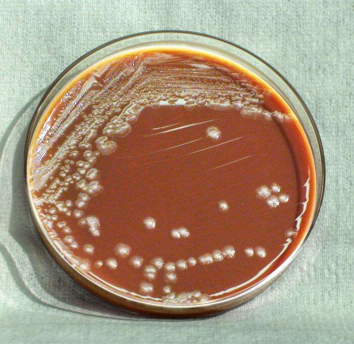
This image depicts a Petri dish containing a MacConkey agar medium, which had been inoculated with Gram-negative Yersinia pestis bacteria. Y. pestis is the pathogen responsible for causing human plague. This was the appearance of the colonial growth after 48 hours of incubation.People usually get plague from being bitten by a rodent flea that is carrying the plague bacterium or by handling an infected animal. Millions of people in Europe died from plague in the Middle Ages, when human homes and places of work were inhabited by flea-infested rats. Today, modern antibiotics are effective against plague, but if an infected person is not treated promptly, the disease is likely to cause illness or death.Created: 2009
-
This image depicts a Petri dish containing a sheeps blood agar (SBA) medium, which had been inoculated with Gram-negative Yersinia pestis bacteria. Y. pestis is the pathogen responsible for causing human plague. This was the appearance of the colonial growth after 48 hours of incubation at 37º C.People usually get plague from being bitten by a rodent flea that is carrying the plague bacterium or by handling an infected animal. Millions of people in Europe died from plague in the Middle Ages, when human homes and places of work were inhabited by flea-infested rats. Today, modern antibiotics are effective against plague, but if an infected person is not treated promptly, the disease is likely to cause illness or death.Created: 2009
-
This image depicts a Petri dish containing a sheeps blood agar (SBA) medium, which had been inoculated with Gram-negative Yersinia pestis bacteria. Y. pestis is the pathogen responsible for causing human plague. This was the appearance of the colonial growth after 48 hours of incubation at 25º C.People usually get plague from being bitten by a rodent flea that is carrying the plague bacterium or by handling an infected animal. Millions of people in Europe died from plague in the Middle Ages, when human homes and places of work were inhabited by flea-infested rats. Today, modern antibiotics are effective against plague, but if an infected person is not treated promptly, the disease is likely to cause illness or death.Created: 2009
-
This image depicts a Petri dish containing a MacConkey agar medium, which had been inoculated with Gram-negative Yersinia pestis bacteria. Y. pestis is the pathogen responsible for causing human plague. This was the appearance of the colonial growth after 24 hours of incubation.People usually get plague from being bitten by a rodent flea that is carrying the plague bacterium or by handling an infected animal. Millions of people in Europe died from plague in the Middle Ages, when human homes and places of work were inhabited by flea-infested rats. Today, modern antibiotics are effective against plague, but if an infected person is not treated promptly, the disease is likely to cause illness or death.Created: 2009
-
This image depicts a Petri dish containing a sheeps blood agar (SBA) medium, which had been inoculated with Gram-negative Yersinia pestis bacteria. Y. pestis is the pathogen responsible for causing human plague. This was the appearance of the colonial growth after 24 hours of incubation at 37º C.People usually get plague from being bitten by a rodent flea that is carrying the plague bacterium or by handling an infected animal. Millions of people in Europe died from plague in the Middle Ages, when human homes and places of work were inhabited by flea-infested rats. Today, modern antibiotics are effective against plague, but if an infected person is not treated promptly, the disease is likely to cause illness or death.Created: 2009
-
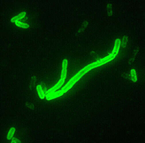
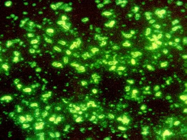
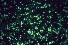
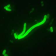
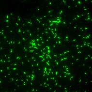
This micrograph was stained using a fluorescent antibody staining technique (FA), which uses the specific conjugated antiserum to Fraction 1 (F1) antigen of Yersinia pestis to identify the antigens present in animal tissues, and appropriate cultures.Created: 1993
-
Description: Deutsch:
Yersinia pestis im Fluoreszenz-Mikroskop mit Fluoreszenz-markiertem Antikörper gegen ein Kapsel-Antigen. Source: Transferred from
de.wikipedia to Commons by
Matthias_M. using
CommonsHelper. (
[1] Herkunft CDC-PHIL). Author: This file is lacking author information.
-
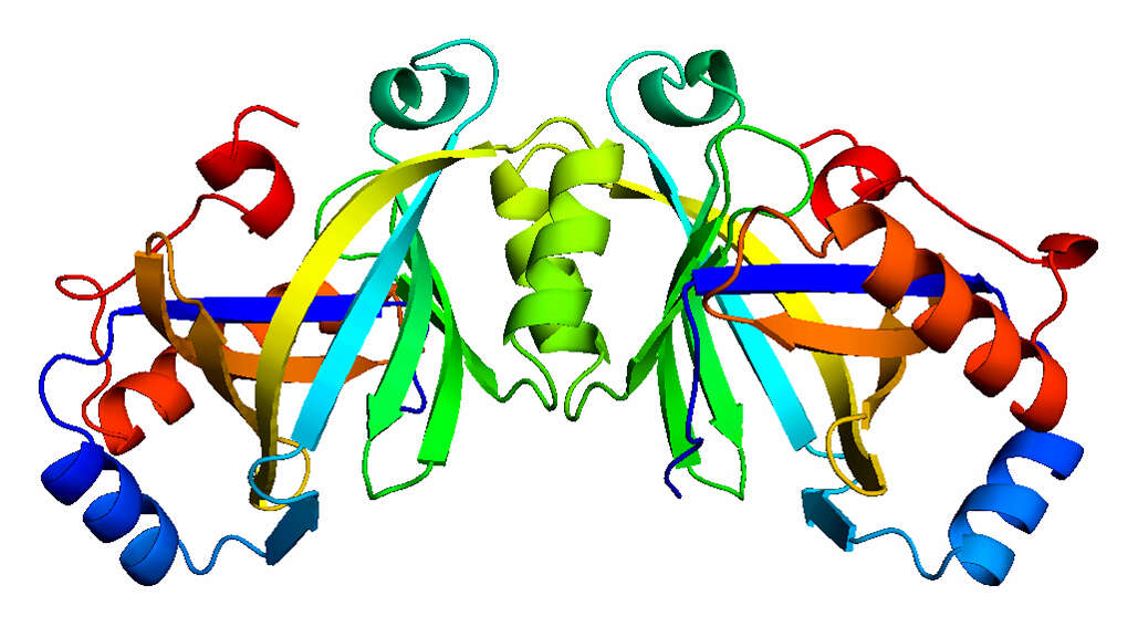
Summary.mw-parser-output table.commons-file-information-table,.mw-parser-output.fileinfotpl-type-information{border:1px solid #a2a9b1;background-color:#f8f9fa;padding:5px;font-size:95%;border-spacing:2px;box-sizing:border-box;margin:0;width:100%}.mw-parser-output table.commons-file-information-table>tbody>tr,.mw-parser-output.fileinfotpl-type-information>tbody>tr{vertical-align:top}.mw-parser-output table.commons-file-information-table>tbody>tr>td,.mw-parser-output table.commons-file-information-table>tbody>tr>th,.mw-parser-output.fileinfotpl-type-information>tbody>tr>td,.mw-parser-output.fileinfotpl-type-information>tbody>tr>th{padding:4px}.mw-parser-output.fileinfo-paramfield{background:#ccf;text-align:right;padding-right:0.4em;width:15%;font-weight:bold}.mw-parser-output.commons-file-information-table+table.commons-file-information-table,.mw-parser-output.commons-file-information-table+div.commons-file-information-table>table{border-top:0;padding-top:0;margin-top:-8px}@media only screen and (max-width:719px){.mw-parser-output table.commons-file-information-table,.mw-parser-output.commons-file-information-table.fileinfotpl-type-information{border-spacing:0;padding:0;word-break:break-word;width:100%!important}.mw-parser-output.commons-file-information-table>tbody,.mw-parser-output.fileinfotpl-type-information>tbody{display:block}.mw-parser-output.commons-file-information-table>tbody>tr>td,.mw-parser-output.commons-file-information-table>tbody>tr>th,.mw-parser-output.fileinfotpl-type-information>tbody>tr>td,.mw-parser-output.fileinfotpl-type-information>tbody>tr>th{padding:0.2em 0.4em;text-align:left;text-align:start}.mw-parser-output.commons-file-information-table>tbody>tr,.mw-parser-output.fileinfotpl-type-information>tbody>tr{display:flex;flex-direction:column}.mw-parser-output.commons-file-information-table+table.commons-file-information-table,.mw-parser-output.commons-file-information-table+div.commons-file-information-table>table{margin-top:-1px}.mw-parser-output.fileinfo-paramfield{box-sizing:border-box;flex:1 0 100%;width:100%}} Description: Ribbon rendering of the structure of AC-IV in Yersina pestis as determined at NIST. The enzyme is a dimer (two identical subunits around a vertical axis), and each of the two subunits forms a central barrel made of eight strands surrounded by short helices. The active site is believed to lie inside the barrel. Credit: NIST Disclaimer: Any mention of commercial products within NIST web pages is for information only; it does not imply recommendation or endorsement by NIST. Use of NIST Information: These World Wide Web pages are provided as a public service by the National Institute of Standards and Technology (NIST). With the exception of material marked as copyrighted, information presented on these pages is considered public information and may be distributed or copied. Use of appropriate byline/photo/image credits is requested. Date: 17 August 2006, 10:39. Source:
Plague Enzyme Structure. Author:
National Institute of Standards and Technology.
-
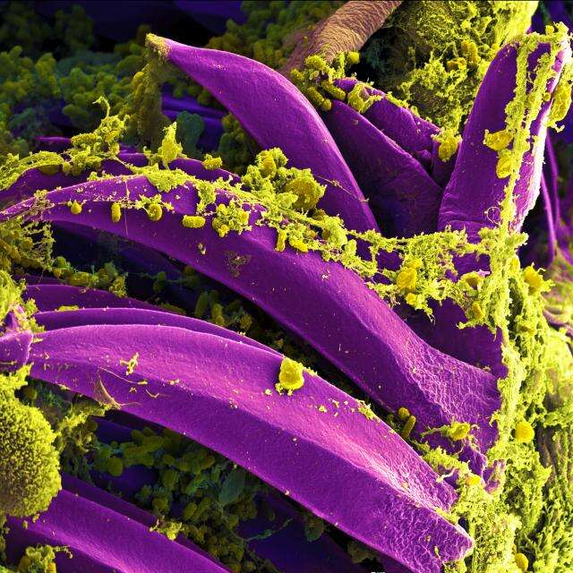
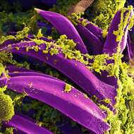
Description: English: Scanning electron micrograph of Yersinia pestis, which causes bubonic plague, on proventricular spines of a Xenopsylla cheopis flea National Institute of Allergy and Infectious Diseases (NIAID). Date: 10 November 2013, 23:47:25. Source: National Institutes of Health (NIH). Author: National Institutes of Health (NIH).
-

Summary Scanning electron microphotograph depicting a mass of Yersinia pestis bacteria (the cause of bubbonic plague) in the foregutte of the flea vector Credit: Rocky Mountain Laboratories, NIAID, NIH
http://www3.niaid.nih.gov/biodefense/Public/Images.htm Text of cropped caption: DD2017 10.0kV X20.0K 1.50µm Licensing[
edit] Public domainPublic domainfalsefalse. : This work is in the
public domain in the United States because it is a
work prepared by an officer or employee of the United States Government as part of that person’s official duties under the terms of
Title 17, Chapter 1, Section 105 of the
US Code. Note: This only applies to original works of the Federal Government and not to the work of any individual
U.S. state,
territory, commonwealth, county, municipality, or any other subdivision. This template also does not apply to postage stamp designs published by the
United States Postal Service since 1978. (See §
313.6(C)(1) of Compendium of U.S. Copyright Office Practices). It also does not apply to certain US coins; see
The US Mint Terms of Use. :.
This file has been identified as being free of known restrictions under copyright law, including all related and neighboring rights.. https://creativecommons.org/publicdomain/mark/1.0/PDMCreative Commons Public Domain Mark 1.0falsefalse
-
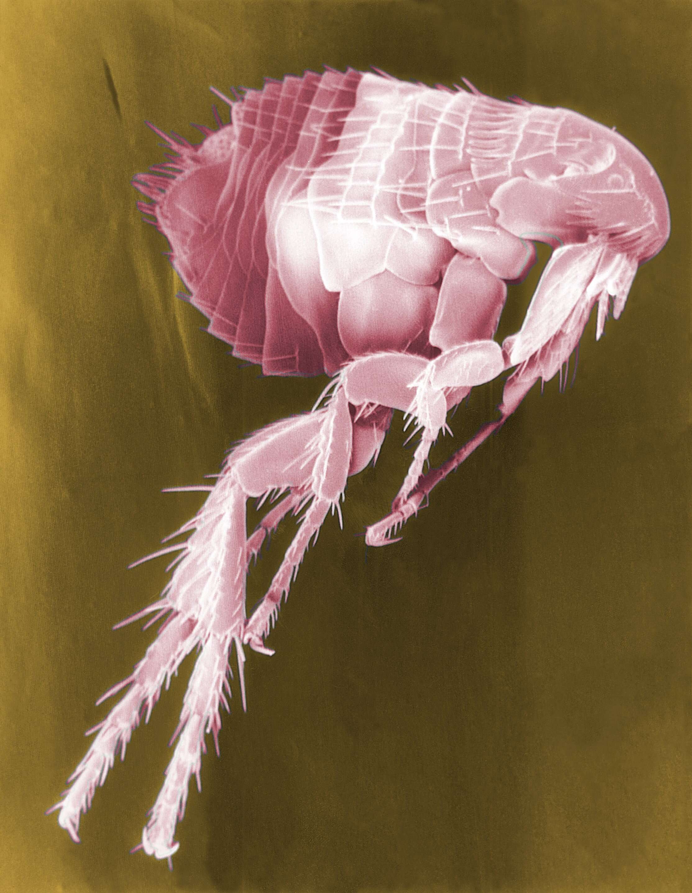
Description[
edit] Description: False colors Scanning Electron Micrograph of a
Flea. Fleas are known to carry a number of diseases that are transferable to human beings through their bites. Included in these infections is the plague, caused by the bacterium Yersinia pestis. Source: : This media comes from the
Centers for Disease Control and Prevention's
Public Health Image Library (PHIL), with identification number
#11436. Note: Not all PHIL images are public domain; be sure to check copyright status and credit authors and content providers.
العربية |
Deutsch |
English |
македонски |
slovenščina |
+/−. Author: CDC/Janice Haney Carr. Permission(
Reusing this file): PD-USGov-HHS-CDC English: None - This image is in the public domain and thus free of any copyright restrictions. As a matter of courtesy we request that the content provider be credited and notified in any public or private usage of this image. Other versions:
Older B&W Version.
-
-
-
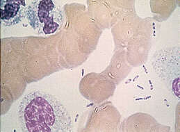
Description: English: Yersinia pestis, Direct Fluorescent Antibody Stain (DFA), 40x Magnification. Positive identification of the bacillus may be facilitated through the application of DFA stain and its affinity for the Y. pestis' capsular antigen. Date: 2002. Source: : This media comes from the
Centers for Disease Control and Prevention's
Public Health Image Library (PHIL), with identification number
#1917. Note: Not all PHIL images are public domain; be sure to check copyright status and credit authors and content providers.
العربية |
Deutsch |
English |
македонски |
slovenščina |
+/−. Author: Photo Credit: Content Providers(s): CDC/ Courtesy of Larry Stauffer, Oregon State Public Health Laboratory. Permission(
Reusing this file): PD-USGov-HHS-CDC (None - This image is in the public domain and thus free of any copyright restrictions. As a matter of courtesy we request that the content provider be credited and notified in any public or private usage of this image.). Other versions:
.
-
Description: English: Re: Simon de Covino Re: Simon de Couvin. Date: 24 October 2019. Source: Own work. Author:
Sean M. Simon.
-
Description: Български: Микроскопска фотография на причинителя на чумата. Картинката е заимствана от
тук, където пък е заимствана от публичния архив на
Centers for Disease Control and Prevention. English:
Yersinia pestis. Source: This file is lacking source information. Please edit this file's description and provide a source. Author: This file is lacking author information.
-
Description: English: This photograph depicts the colonial morphology displayed by Gram-negative Yersinia pestis bacteria, which was grown on a medium of chocolate agar, for a 72 hour time period, at a temperature of 25°C. Note that PHIL 12470 displays a closer view of these Y. pestis bacterial colonies. See also PHIL 12468 for the appearance of Y. pestis colonies grown on the same medium, and at the same time temperature, but for a lesser time period (48hr); PHIL 12471 displaying growth on the same medium, but for a lesser time period (48hr), and at a greater temperature (37°C); PHIL 12472 displaying growth on the same medium, for the same amount of time, but at a greater temperature (37°C); PHIL 12473 displaying Y. pestis colonies grown on the same medium and temperature, but at one-third the time (24hr). Date: 2010. Source: : This media comes from the
Centers for Disease Control and Prevention's
Public Health Image Library (PHIL), with identification number
#12469. Note: Not all PHIL images are public domain; be sure to check copyright status and credit authors and content providers.
العربية |
Deutsch |
English |
македонски |
slovenščina |
+/−. Author: Department of Health and Human Services.
-
-
Description: English: Yersinia pestis. Date: 30 April 2008. Source: Own work. Author: A.Myasnikov for Wiki.
-


