Browse Trichoplax adhaerens genome and functional genomics using the JGI browser.
Pearse and Voigt (2007) report that in their sampling using glass slides, certain general types of organisms were often found in association with placozoans, including several kinds of sessile ciliates (solitary and colonial vorticellids as well as folliculinids); spirorbid and other serpulid polychaetes; and, in smaller numbers, free-living loxosomatid kamptozoans (entoprocts). Placozoans have been observed capturing and eating ciliates in a culture dish. Wild placozoans are probably opportunistic grazers and scavengers on organic detritus and on algae and bacteria in biofilms covering a diversity of substrates. (Pearse and Voigt 2007 and references therein)
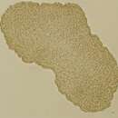
Trichoplax adhaerens, the only described species in the phylum Placozoa, is the structurally simplest metazoan (multicellular animal). It is quite small (just 2 to 3 mm in diameter) and consists of several thousand cells arranged as a double-layered plate. It lacks anterior-posterior polarity and symmetry. However, the cells of the upper and lower layers differ in shape and there is a consistent dorsal-ventral orientation of the body relative to the substrate. Trichoplax moves by ciliary gliding, changing its shape along the edges as it moves, like an amoeba. Very small (presumably young) individuals can swim, but larger individuals crawl. It appears that Trichoplax feeds by phagocytosis of organic detritus. (Brusca and Brusca 2003) Although the discovery and description of Trichoplax adhaerens in 1883 (in saltwater tanks in Austria) sparked controversy among zoologists about what its significance might be with respect to inferring the characteristics of early metazoans, interest faded away as the (incorrect) idea took hold that these enigmatic organisms were simply abnormal larvae of hydrozoan cnidarians. Although strong evidence against this view was published in 1912 and 1914, there was no more discussion of Trichoplax in the zoological literature--nor citation of the key critiques of 1912 and 1914-- for more than half a century (although the misconception that it was a cnidarian was repeated in textbooks). Beginning in the 1960s, several researchers re-focused attention on Trichoplax, demonstrating that it is an adult form of a new phylum dubbed, in 1971, Placozoa. It became clear that placozoans could be found worldwide in the shallow waters of subtropical and tropical regions (Pearse 1989; Eitel and Schierwater 2010). (Syed and Schierwater 2002) Recent data all indicate that the Placozoa represent one of the earliest branching lineages in the metazoan tree, but the exact placement of this branch remains uncertain (Schierwater et al. 2009; Ball and Miller 2010 and references therein).
Pearse and Voigt (2007) summarized all the sites where placozoans were known to have been collected up to the time of their publication. Based on current knowledge, Eitel and Schierwater (2010) predict that most placozoans are found between the equator and 20 degrees North.
Dellaporta et al. (2006) report on the complete mitochondrial genome of Trichoplax adhaerens, which they note is the largest known metazoan mtDNA genome--at 43,079 bp, it is more than twice the size of the typical metazoan mtDNA. Srivastava et al. (2008) present a preliminary analysis of the genome of Trichoplax adhaerens, which is among the smallest known metazoan genomes at about 98 million base pairs.
Placozoans have been found only in shallow, nearshore, tropical and subtropical marine environments between approximately 30 degrees north and south of the Equator. Within these habitats, they tend to be absent from areas of high current and from bare sand bottoms. Because they are very sensitive to reductions in salinity, they will presumably not be found in fresh or brackish water or seasonally in areas that are subjected to heavy monsoon rains. (Ball and Miller 2010 and references therein; Eitel and Schierwater 2010) They are often found on mangrove tree roots, reefs, boat docks in the eulittoral and littoral zone, and around stony beaches (Eitel and Schierwater 2010). They can be abundant on coral reefs. Although placozoans have long been viewed as benthic organisms, Pearse and Voigt (2007) had more success collecting them on glass slides suspended 20 to 60 cm above the bottom than on glass slides situated on the bottom itself.
The placozoan Trichoplax adhaerens is a small (1 to 2 mm) ciliated disc-shaped microscopic marine animal. The thin body consists only of an epithelium and an internal mesenchyme. The epithelium has two regions: an upper free, or dorsal, epithelium of cover cells and a lower attached, or ventral, epithelium of cylinder and gland cells. Some differences between the central and marginal areas in the body have also been reported. When the densely ciliated ventral epithelium is in contact with the substrate, the animals display a gliding or creeping locomotion. When the animal is feeding, its shape changes periodically. (Ueda et al. 1999; Maruyama 2004 and references therein; Srivastava et al. 2008)
When large or stressed, placozoans can undergo binary fission (split into two individuals--sometimes three!) or bud to produce numerous multicellular flagellated "swarmers", which are capable of regenerating the amoeboid stage. However, this is not the complete life cycle, as there is also evidence of sexual reproduction. (Brusca and Brusca 2003; Ball and Miller 2010 and references therein) Sexual reproduction has not been observed in culture, but putative oocyte formation in degenerating animals is seen routinely. Population genetic analyses demonstrate allelic variation and evidence for genetic recombination in animals in the wild that is consistent with sex. (Signorovitch et al. 2005; Srivastava et al. 2008)
Trichoplax adhaerens is the only described species in the phylum Placozoa, but recent work (Eitel and Schierwater 2010 and references therein) suggests that there may be many unrecognized placozoan species.
Trichoplax adhaerens is one of the four named species in the phylum Placozoa. The others are Hoilungia hongkongensis, Polyplacotoma mediterranea and Cladtertia collaboinventa. Placozoa is a basal group of multicellular animals, possible relatives of Cnidaria.[2] Trichoplax are very flat organisms around a millimetre in diameter, lacking any organs or internal structures. They have two cellular layers: the top epitheloid layer is made of ciliated "cover cells" flattened toward the outside of the organism, and the bottom layer is made up of cylinder cells that possess cilia used in locomotion, and gland cells that lack cilia.[3] Between these layers is the fibre syncytium, a liquid-filled cavity strutted open by star-like fibres.
Trichoplax feed by absorbing food particles—mainly microbes—with their underside. They generally reproduce asexually, by dividing or budding, but can also reproduce sexually. Though Trichoplax has a small genome in comparison to other animals, nearly 87% of its 11,514 predicted protein-coding genes are identifiably similar to known genes in other animals.
Trichoplax was discovered in 1883 by the German zoologist Franz Eilhard Schulze, in a seawater aquarium at the Zoological Institute in Graz, Austria. The generic name is derived from the classical Greek θρίξ (thrix), "hair", and πλάξ (plax), "plate". The specific epithet adhaerens is Latin meaning "adherent", reflecting its propensity to stick to the glass slides and pipettes used in its examination.[4]
Although from the very beginning most researchers who studied Trichoplax in any detail realized that it had no close relationship to other animal phyla, the zoologist Thilo Krumbach published a hypothesis that Trichoplax is a form of the planula larva of the anemone-like hydrozoan Eleutheria krohni in 1907. Although this was refuted in print by Schulze and others, Krumbach's analysis became the standard textbook explanation, and nothing was printed in zoological journals about Trichoplax until the 1960s. In the 1960s and 1970s a new interest among researchers led to acceptance of Placozoa as a new animal phylum. Among the new discoveries was study of the early phases of the animals' embryonic development and evidence that the animals that people had been studying are adults, not larvae. This newfound interest also included study of the organism in nature (as opposed to aquariums).[5]

Trichoplax generally has a thinly flattened, plate-like body in cross-section around half a millimetre, occasionally up to two or three millimetres. The body is usually only about 25 µm thick. These colorlessly gray organisms are so thin they are transparent when illuminated from behind, and in most cases are barely visible to the naked eye. Like the single-celled amoebae, which they superficially resemble, they continually change their external shape. In addition, spherical phases occasionally form. These may facilitate movement to new habitats.
Trichoplax lacks tissues and organs; there is also no manifest body symmetry, so it is not possible to distinguish anterior from posterior or left from right. It is made up of a few thousand cells of six types in three distinct layers: dorsal epithelia cells and ventral epithelia cells, each with a single cilium ("monociliate"), ventral gland cells, syncytial fiber cells, lipophils, and crystal cells (each containing a birefringent crystal, arrayed around the rim). Lacking sensory and muscle cells, it moves using cilia on its external surface.[6]
There are no neurons present, but in the absence of a nervous system the animal uses short chains of amino acids known as peptides for cell communication, in a manner resembling the way animals with neurons use neuropeptides for the same purpose. Individual cells contain and secrete a variety of small peptides, made up of between four and 20 amino acids, which are detected by neighbouring cells. Each peptide can be used individually to send a signal to other cells, but also sequentially or together in different combinations, creating a huge number a different types of signals. This allows for a relatively complex behavioural repertoire, including behaviours such as "crinkling", turning, flattening, and internal "churning".[7]
Both structurally and functionally, it is possible to distinguish a back or dorsal side from a belly or ventral side in Trichoplax adhaerens. Both consist of a single layer of cells coated on the outside with slime and are reminiscent of epithelial tissue, primarily due to the junctions—belt desmosomes—between the cells. In contrast to true epithelium, however, the cell layers of the Placozoa possess no basal lamina, which refers to a thin layer of extracellular material underlying epithelium that stiffens it and separates it from the body's interior. The absence of this structure, which is otherwise to be found in all animals except the sponges, can be explained in terms of function: a rigid separating layer would make the amoeboid changes in the shape of Trichoplax adhaerens impossible. Instead of an epithelium, therefore, we speak of an epitheloid in the Placozoa.
A mature individual consists of up to a thousand cells that can be divided into four different cell types. The monociliated cells of the dorsal epitheloid are flattened and contain lipid bodies. The cells on the ventral side likewise possess a single cilium, while their elongated columnar shape, with a small cross section at the surface, packs them very closely together, causing the cilia to be very closely spaced on the ventral side and to form a ciliated "crawling sole". Interspersed among these ventral epithlioid cells are unciliated gland cells thought to be capable of synthesizing digestive enzymes.
Between the two layers of cells is a liquid-filled interior space, which, except for the immediate zones of contact with the ventral and dorsal sides, is pervaded by a star-shaped fibre syncytium: a fibrous network that consists essentially of a single cell but contains numerous nuclei that, while separated by internal crosswalls (septa), do not have true cell membranes between them. Similar structures are also found in the sponges (Porifera) and many fungi.
On both sides of the septa are liquid-filled capsules that cause the septa to resemble synapses, i.e. nerve-cell junctions that occur in fully expressed form only in animals with tissues (Eumetazoa). Striking accumulations of calcium ions, which may have a function related to the propagation of stimuli, likewise suggest a possible role as protosynapses. This view is supported by the fact that fluorescent antibodies against cnidarian neurotransmitters, i.e. precisely those signal carriers that are transferred in synapses, bind in high concentrations in certain cells of Trichoplax adhaerens, and thus indicate the existence of comparable substances in the Placozoa. The fibre syncytium also contains molecules of actin and probably also of myosin, which occur in the muscle cells of eumetazoans. In the placozoans, they ensure that the individual fibres can relax or contract and thus help determine the animals' shape.
In this way, the fibre syncytium assumes the functions of nerve and muscle tissues. Moreover, at least a portion of digestion occurs here. On the other hand, no gelatinous extracellular matrix exists of the kind observed, in mesoglea, in cnidarians and ctenophores.
Pluripotent cells, which can differentiate into other cell types, have not yet been demonstrated unambiguously in T. adhaerens, in contrast to the case of the Eumetazoa. The conventional view is that dorsal and ventral epithelioid cells arise only from other cells of the same type.
The Trichoplax genome contains about 98 million base pairs and 11,514 predicted protein-coding genes.[8]
All nuclei of placozoan cells contain six pairs of chromosomes that are only about two to three micrometres in size. Three pairs are metacentric, meaning that the centromere, the attachment point for the spindle fibers in cell division, is located at the center, or acrocentric, with the centromere at an extreme end of each chromosome. The cells of the fiber syncytium can be tetraploid, i.e. contain a quadruple complement of chromosomes.
A single complement of chromosomes in Trichoplax adhaerens contains a total of fewer than fifty million base pairs and thus forms the smallest animal genome; the number of base pairs in the intestinal bacterium Escherichia coli is smaller by a factor of only ten.
The genetic complement of Trichoplax adhaerens has not yet been very well researched; it has, however, already been possible to identify several genes, such as Brachyury and TBX2/TBX3, which are homologous to corresponding base-pair sequences in eumetazoans. Of particular significance is Trox-2, a placozoan gene known under the name Cnox-2 in cnidarians and as Gsx in the bilaterally symmetrical Bilateria. As a homeobox or Hox gene it plays a role in organization and differentiation along the axis of symmetry in the embryonic development of eumetazoans; in cnidarians, it appears to determine the position of mouth-facing (oral) and opposite-facing (aboral) sides of the organism. Since placozoans possess no axes of symmetry, exactly where the gene is transcribed in the body of Trichoplax is of special interest. Antibody studies have been able to show that the gene's product occurs only in the transition zones of the dorsal and ventral sides, perhaps in a fifth cell type that has not yet been characterized. It is not yet clear whether these cells, contrary to traditional views, are stem cells, which play a role in cell differentiation. In any case, Trox-2 can be considered a possible candidate for a proto-Hox gene, from which the other genes in this important family could have arisen through gene duplication and variation.
Initially, molecular-biology methods were applied unsuccessfully to test the various theories regarding Placozoa's position in the Metazoa system. No clarification was achieved with standard markers such as 18S rDNA/RNA: the marker sequence was apparently "garbled", i.e. rendered uninformative as the result of many mutations. Nevertheless, this negative result supported the suspicion that Trichoplax might represent an extremely primitive lineage of metazoans, since a very long period of time had to be assumed for the accumulation of so many mutations.
Of the 11,514 genes identified in the six chromosomes of Trichoplax, 87% are identifiably similar to genes in cnidarians and bilaterians. In those Trichoplax genes for which equivalent genes can be identified in the human genome, over 80% of the introns (the regions within genes that are removed from RNA molecules before their sequences are translated in protein synthesis) are found in the same location as in the corresponding human genes. The arrangement of genes in groups on chromosomes is also conserved between the Trichoplax and human genomes. This contrasts to other model systems such as fruit flies and soil nematodes that have experienced a paring down of non-coding regions and a loss of the ancestral genome organizations.[9]
The phylogenetic relationship between Trichoplax and other animals has been debated for some time. A variety of hypotheses have been advanced based on the few morphological characteristics of this simple organism that could be identified. More recently, a comparison of the Trichoplax mitochondrial genome suggested that Trichoplax is a basal metazoan—less closely related to all other animals including sponges than they are to each other.[10] This implies that the Placozoa would have arisen relatively soon after the evolutionary transition from unicellular to multicellular forms. But an even more recent analysis of the much larger Trichoplax nuclear genome instead supports the hypothesis that Trichoplax is a basal eumetazoan, that is, more closely related to Cnidaria and other animals than any of those animals are to sponges.[8] This is consistent with the presence in Trichoplax of cell layers reminiscent of epithelial tissue (see above).
Trichoplax was first discovered on the walls of a marine aquarium, and is rarely observed in its natural habitat.[11] Trichoplax has been collected, among other places, in the Red Sea, the Mediterranean, and the Caribbean, off Hawaii, Guam, Samoa, Japan, Vietnam, Brazil, and Papua New Guinea, and on the Great Barrier Reef off the east coast of Australia.[12]
Field specimens tend to be found in the coastal tidal zones of tropical and subtropical seas, on such substrates as the trunks and roots of mangroves, shells of molluscs, fragments of stony corals or simply on pieces of rock. One study was able to detect seasonal population fluctuations, the causes of which have not yet been deduced.
Trichoplax adhaerens feeds on small algae, particularly on green algae (Chlorophyta) of the genus Chlorella, cryptomonads (Cryptophyta) of the genera Cryptomonas and Rhodomonas, and blue-green bacteria (Cyanobacteria) such as Phormidium inundatum, but also on detritus from other organisms. In feeding, one or several small pockets form around particles of nutrients on the ventral side, into which digestive enzymes are released by the gland cells; the organisms thus develop a temporary "external stomach", so to speak. The enclosed nutrients are then taken up by pinocytosis ("cell-drinking") by the ciliated cells located on the ventral surface.
Entire single-celled organisms can also be ingested through the upper epitheloid (that is, the "dorsal surface" of the animal). This mode of feeding could be unique in the animal kingdom: the particles, collected in a slime layer, are drawn through the intercellular gaps (cellular interstices) of the epitheloid by the fibre cells and then digested by phagocytosis ("cell-eating"). Such "collecting" of nutrient particles through an intact tegument is only possible because some "insulating" elements (specifically, a basal lamina under the epitheloid and certain types of cell-cell junctions) are not present in the Placozoa.
Not all bacteria in the interior of Placozoa are digested as food: in the endoplasmic reticulum, an organelle of the fibre syncytium, bacteria are frequently found that appear to live in symbiosis with Trichoplax adhaerens.[13]
Placozoa can move in two different ways on solid surfaces: first, their ciliated crawling sole lets them glide slowly across the substrate; second, they can change location by modifying their body shape, as an amoeba does. These movements are not centrally coordinated, since no muscle or nerve tissues exist. It can happen that an individual moves simultaneously in two different directions and consequently divides into two parts.[14]
It has been possible to demonstrate a close connection between body shape and the speed of locomotion, which is also a function of available food:
Since the transition is not smooth but happens abruptly, the two modes of extension can be very clearly separated from one another. The following is a qualitative explanation of the animal's behavior:
The actual direction in which Trichoplax moves each time is random: if we measure how fast an individual animal moves away from an arbitrary starting point, we find a linear relationship between elapsed time and mean square distance between starting point and present location. Such a relationship is also characteristic of random Brownian motion of molecules, which thus can serve as a model for locomotion in the Placozoa.
Small animals are also capable of swimming actively with the aid of their cilia. As soon as they come into contact with a possible substrate, a dorsoventral response occurs: the dorsal cilia continue to beat, whereas the cilia of ventral cells stop their rhythmic beating. At the same time, the ventral surface tries to make contact with the substrate; small protrusions and invaginations, the microvilli found on the surface of the columnar cells, help in attaching to the substrate via their adhesive action.
Using Trichoplax adhaerens as a model, were described 0.02–0.002 Hz oscillations in locomotory and feeding patterns as evidence of complex multicellular integration; and showed their dependence on the endogenous secretion of signal molecules. Evolutionary conserved low-molecular-weight transmitters (glutamate, aspartate, glycine, GABA, and ATP) acted as coordinators of distinct locomotory and feeding patterns. Specifically, L-glutamate induced and partially mimicked endogenous feeding cycles, whereas glycine and GABA suppressed feeding. ATP-modified feeding is complex, first causing feeding-like cycles and then suppressing feeding. Trichoplax locomotion was modulated by glycine, GABA, and, surprisingly, by animals’ own mucus trails. Mucus triples locomotory speed compared to clean substrates. Glycine and GABA increased the frequency of turns. [15]
A notable characteristic of the Placozoa is that they can regenerate themselves from extremely small groups of cells. Even when large portions of the organism are removed in the laboratory, a complete animal develops again from the remainder. It is also possible to rub Trichoplax adhaerens through a strainer in such a manner that individual cells are not destroyed but are separated from one another to a large extent. In the test tube they then find their way back together again to form complete organisms. If this procedure is performed on several previously strained individuals simultaneously, the same thing occurs. In this case, however, cells that previously belonged to a particular individual can suddenly show up as part of another.
The Placozoa normally propagate asexually, dividing down the middle to produce two (or sometimes, three) roughly equal-sized daughters. These remain loosely connected for a while after fission. More rarely, budding processes are observed: spherules of cells separate from the dorsal surface; each of these combines all known cell types and subsequently grows into an individual on its own.
Sexual reproduction is thought to be triggered by excessive population density. As a result, the animals absorb liquid, begin to swell, and separate from the substrate so that they float freely in the water. In the protected interior space, the ventral cells form an ovum surrounded by a special envelope, the fertilisation membrane; the ovum is supplied with nutrients by the surrounding syncytium, allowing energy-rich yolk to accumulate in its interior. Once maturation of the ovum is complete, the rest of the animal degenerates, liberating the ovum itself. Small, unciliated cells that form at the same time are interpreted to be spermatozoa. It has not yet been possible to observe fertilisation itself; the existence of the fertilisation membrane is currently taken to be evidence, however, that it has taken place.
Putative eggs have been observed, but they degrade, typically at the 32–64 cell stage. Neither embryonic development nor sperm have been observed. Despite lack of observation of sexual reproduction in the lab, the genetic structure of the populations in the wild is compatible with the sexual reproduction mode, at least for species of the analysed genotype H5.[16]
Usually even before its liberation, the ovum initiates cleavage processes in which it becomes completely pinched through at the middle. A ball of cells characteristic of animals, the blastula, is ultimately produced in this manner, with a maximum of 256 cells. Development beyond this 256-cell stage has not yet been observed.[17]
Trichoplax lack a homologue of the Boule protein that appears to be ubiquitous and conserved in males of all species of other animals tested.[18] If its absence implies the species has no males, then perhaps its "sexual" reproduction may be a case of the above-described process of regeneration, combining cells from two separate organisms into one.
Due to the possibility of its cloning itself by asexual propagation without limit, the life span of Placozoa is infinite; in the laboratory, several lines descended from a single organism have been maintained in culture for an average of 20 years without the occurrence of sexual processes.
Long ignored as an exotic, marginal phenomenon, Trichoplax adhaerens is today viewed as a potential biological model organism. In particular, research is needed to determine how a group of cells that cannot be considered full-fledged epithelial tissue organizes itself, how locomotion and coordination occur in the absence of true muscle and nerve tissue, and how the absence of a concrete body axis affects the animal's biology. At the genetic level, the way in which Trichoplax adhaerens protects against damage to its genome needs to be studied, particularly with regard to the existence of special DNA-repair processes. T. adhaerens can tolerate high levels of radiation damage that are lethal to other animals.[19] Tolerance to X-ray exposure was found to depend on expression of genes involved in DNA repair and apoptosis including the gene Mdm2.[19] Complete decoding of the genome should also clarify the placozoans' place in evolution, which continues to be controversial.
Its ability to fight cancer through a combination of aggressive DNA repair and ejection of damaged cells makes it a promising organism for cancer research.[20]
In addition to basic research, this animal could also be suitable for studying wound-healing and regeneration processes; as yet unidentified metabolic products should be researched. Finally, Trichoplax adhaerens is also being considered as an animal model for testing compounds and antibacterial drugs.[21]
Francesco Saverio Monticelli described another species in 1893, which he found in the waters around Naples, naming it Treptoplax reptans. However, it has not been observed since 1896, and most zoologists today doubt its existence.
Significant genetic differences have been observed between collected specimens matching the morphological description of T. adhaerens, suggesting that it may be a cryptic species complex. At least eight distinct genotypes (marked from H1 to H8) have been observed.[22]
Because great genetic differences often occur between representatives of Trichoplax adhaerens, differences that in other taxa would result in their being spread among different genera, it is currently unclear whether the single species, based on morphological criteria, does not actually correspond to a group of cryptospecies, i.e. species that are not outwardly distinguishable from one another. Distribution of the genetic variants is not a function of geography: some variants are found in multiple regions (e.g. Pacific, Caribbean and Red Sea). At the same time, very different genetic variants can be isolated from the same habitat.
{{cite journal}}: CS1 maint: multiple names: authors list (link) {{cite web}}: CS1 maint: url-status (link) {{cite journal}}: CS1 maint: multiple names: authors list (link) Trichoplax adhaerens is one of the four named species in the phylum Placozoa. The others are Hoilungia hongkongensis, Polyplacotoma mediterranea and Cladtertia collaboinventa. Placozoa is a basal group of multicellular animals, possible relatives of Cnidaria. Trichoplax are very flat organisms around a millimetre in diameter, lacking any organs or internal structures. They have two cellular layers: the top epitheloid layer is made of ciliated "cover cells" flattened toward the outside of the organism, and the bottom layer is made up of cylinder cells that possess cilia used in locomotion, and gland cells that lack cilia. Between these layers is the fibre syncytium, a liquid-filled cavity strutted open by star-like fibres.
Trichoplax feed by absorbing food particles—mainly microbes—with their underside. They generally reproduce asexually, by dividing or budding, but can also reproduce sexually. Though Trichoplax has a small genome in comparison to other animals, nearly 87% of its 11,514 predicted protein-coding genes are identifiably similar to known genes in other animals.
